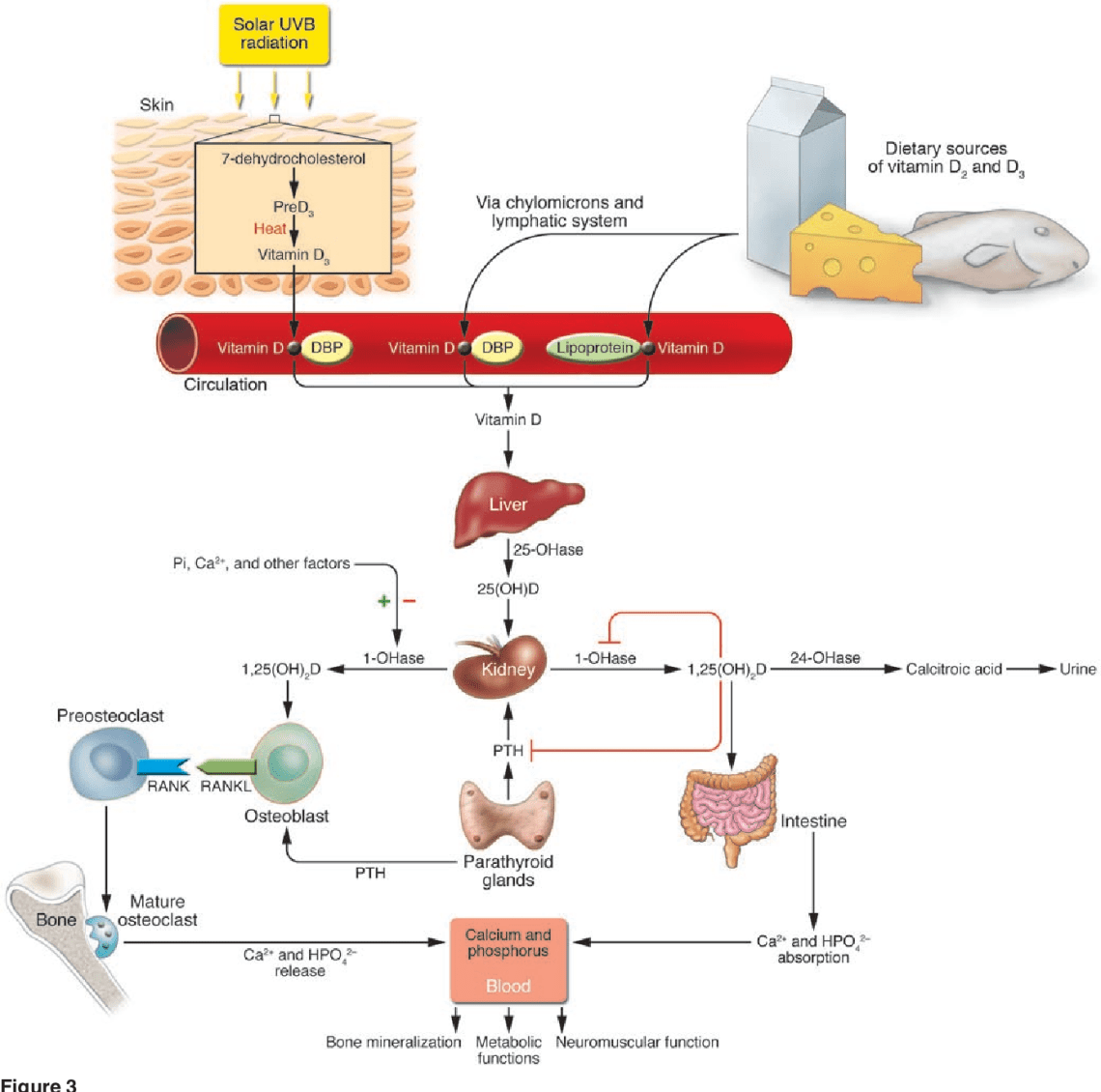
Vitamin D3 thiab calcium deficiency (rickets, hypocalcification, osteopenia)
Cov tsos mob: soft or crooked shell Vaub kib: dej thiab av Kev kho mob: can be cured on its own, running is not treated
This is the most common group of diseases when keeping turtles in captivity. Rickets is a special case of calcium imbalance diseases. Diseases of this group can occur in different forms, but in all cases it is associated to one degree or another with a decrease in the concentration of calcium in the bone tissue.
Osteopenia is a collective term for abnormally low bone mass. There are three types of osteopenic lesions: osteoporosis (simultaneous loss of organic matrix and minerals), osteomalacia (insufficient bone mineralization), fibrocystic osteitis (increased resorption of the main bone substance and its replacement with fibrous tissue).
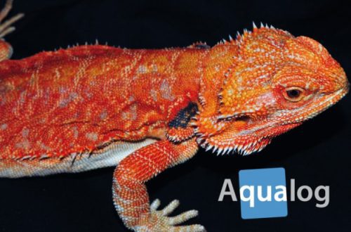
Normally, a turtle’s shell should be even, without bumps and dips, approximately uniform in color, domed for terrestrial and elongated streamlined for aquatic.
")
")
")
Qhov laj thawj:
When turtles are fed with feed mixtures that are not enriched with calcium and vitamin D3, as well as in the absence of natural or artificial ultraviolet radiation, all turtles, both young and adults, develop a pattern of calcium leaching from the body. Some foods also help flush out calcium from the body, such as white cabbage.
Cov tsos mob:
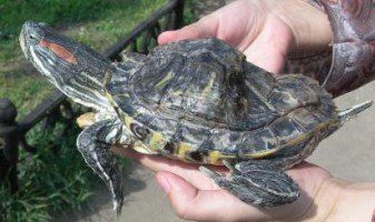
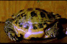
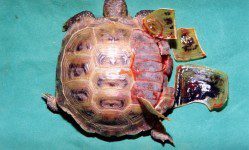
Young water turtles: the shell becomes soft and, as it were, cramped for the turtle; Normally, in young turtles, the shell should harden by the end of the first year of life. Young tortoises: pyramidal growth of the shell and curvature of the limbs.
adult turtles: failure in the posterior third of the carapace, which cannot withstand the pressure of the muscles of the pelvic girdle. The entire shell becomes lighter and flatter. The bony scutes in the area of the bridge between the carapace and the plastron grow (here the bones are more spongy) and the distance between the upper and lower carapace increases. The carapace, especially the plastron, may be soft on palpation. The shell can grow uncontrollably, and the turtle takes on a kind of spherical shape.
Old turtles: the shell usually does not become soft, but becomes very light and resembles plastic. The turtle seems “empty” inside (due to the thickening and porosity of the bone plates). However, the total weight of the turtle may remain within the normal range due to the development of edema in the body cavity.
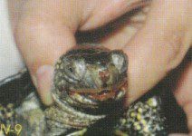
Tsis tas li ntawd, muaj: spontaneous fractures of the limbs, bleeding, prolapse of the cloaca, the turtle cannot lift the body when walking and, as it were, floats, touching the ground with its plastron; the turtle moves only on its front legs – due to weakness or paresis of the hind legs; aquatic turtles are not able to get out on their “raft” and, if a gentle shore is not built in the terrarium, they can drown; the beak is more like a duck (the shape of the bite changes irreversibly, which will no longer allow the turtle to eat the roughage it needs). In the last stage, death can occur from diffuse hemorrhage, acute heart failure and pulmonary edema. When calcium in the diet is normal and phosphorus is in excess, edema and fluid accumulation under the plastron shields may develop, but bleeding is usually absent. Many other diseases can cause similar symptoms, so the turtle should be examined by a veterinarian who will do tests and determine the amount of calcium and phosphorus in the body.
With osteopenia, paresis or weakness of the hind limbs, impaired flotation and regurgitation of mucus from the stomach are possible, i.e. mimic pneumonia in terms of symptoms. There may be problems with breathing (it becomes hoarse and heavy), the skin is clammy, yellow sticky flakes in the folds of the skin.
")
")
XIM: Cov kev kho mob ntawm qhov chaw tuaj yeem ua tau dhau caij nyoog! Tus vaub kib tuaj yeem muaj ntau yam kab mob ib zaug, thiab ntau yam kab mob nyuaj rau kev kuaj mob yam tsis muaj kev kuaj thiab tshuaj xyuas los ntawm kws kho tsiaj, yog li ua ntej pib kho tus kheej, hu rau tsev kho tsiaj nrog tus kws kho tsiaj uas ntseeg siab, lossis peb tus kws kho tsiaj hauv lub rooj sab laj.
Txoj Kev Kho Mob
When examining rickety turtles, increased caution is necessary – bone fractures and deformation of soft organs are possible. The fall of such turtles, even from a small height, is fraught with serious injuries. Any diagnosis in particular “rickets” should be made by a veterinarian. Softening of the shell may be associated with renal failure, hyperparathyroidism, alimentary osteodystrophy, classic “rickets” (lack of vitamin D3), etc.
Rickets I-II stage (the limbs work normally, there are no systemic symptoms: bleeding, swelling and paresis).
- Enter Calcium gluconate (10% solution) at a dosage of 1 ml/kg or Calcium Borgluconate (20% solution) at a dosage of 0,5 ml/kg, intramuscularly or subcutaneously (up to 0.02 intramuscularly, more – s / c ), every 24 or 48 hours depending on the degree of rickets for 2-14 days.
- Drink Panangin (potassium and magnesium) at 1 ml / kg every other day for 10 days. Panangin helps calcium to go to the bones and shell, and not to the joints.
- If the turtle eats on its own, sprinkle 1-2 times a week on food or in food calcium top dressing for reptiles (or crushed cuttlefish shell – sepia).
- The turtle should be exposed to active UV light (ultraviolet lamp for reptiles 10% UVB). Daily for 10-12 hours.
- It is necessary to adjust the diet of aquatic turtles by adding more calcium-containing foods to it. For aquatic turtles, these are Reptomin (Tetra), shelled shrimp, small-boned fish, and small shelled snails.
Treatment will require 2 to 8 weeks.
Rickets III-IV stages (note paresis of the limbs and intestines, spontaneous fractures and bleeding, anorexia, lethargy and shortness of breath).
Treatment is prescribed and carried out by a veterinarian. Treatment takes at least 2 – 3 months. During the first year, it is necessary to monitor the diet and, if possible, the biochemical parameters of the blood.
*Calcium injections – there are several ways to administer calcium – intramuscular and subcutaneous. In each case, this issue should be decided by the attending physician or a specialist consulting on the forum.
Rau kev kho mob koj yuav tsum yuav:
- Calcium Borgluconate Solution | 1 vial | veterinary pharmacy or Calcium Gluconate Solution | 1 vial | human pharmacy
- Panangin | 1 vial | human pharmacy
- Syringe 1 ml | 1 daim | tib neeg lub tsev muag tshuaj
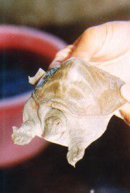
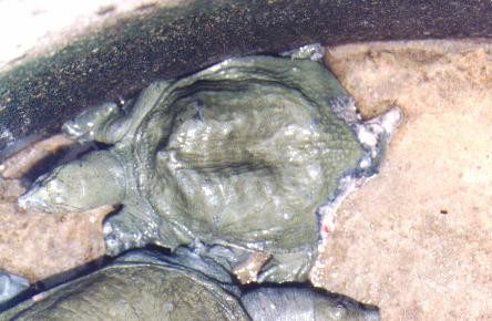
Also in turtles, kyphosis (congenital or acquired) is possible: In wild turtles, kyphosis is a congenital condition. It sometimes appears in various species and is especially pronounced in three-clawed ones, when the turtle becomes similar to a sombrero.
| and lordosis (“collapsing” back)
|


Koj Yuav Tsis tas li ntawd Xav
Euthanasia ntawm cov tsiaj reptiles thiab amphibians
Kev nyab xeeb yooj yim thaum ua haujlwm nrog terrarium thiab terrarium tsiaj